Group of attendees practice Active Release Techniques on each other during the hands-on portion of the webinar.

Nerve

Nerve combines all ART nerve protocols into a single seminar. This is an advanced seminar requiring mastery of Upper Extremity, Lower Extremity, and Spine Level 1 skills in order to successfully complete the seminar, becoming credentialed as an ART Nerve Certified Provider™. Attendees will be trained to identify and treat pain, disorder, and discomfort originating from nerve entrapment, damage or injury, anywhere in the body.

Prerequisites

Nerve attendees must be ART Full Body Level 1 certified, meaning successful completion of ART Upper Extremity Level 1, Lower Extremity Level 1, and Spine Level 1 is required prior to enrolling in the seminar.

The Nerve Seminar was full of fantastic information, from the research and clinical aspect. It inspired me to take a more in-depth look at cases that walk into our office on a daily basis that often seem at first like just a muscular issue. Taking the time to palpate the nerves, knowing how and when to look for a problem with a nerve that isn’t the “classic” nerve pain, enabled me to more effectively treat these conditions in my patients with great skill and precision.

Scott Keller
ART Certified Provider and Instructor

The new Nerve seminar helped me in many ways. It was a great consolidation of the nerve protocols spread out over the Level 1 seminars, clarified concepts of terminal vs local vs long tract which is very helpful in understanding the different nerve protocols, and the treatment process for long tract nerves has been simplified. I finally feel confident using it on my patients!

Joe Scota
ART Certified Provider and Instructor

Nerve is a breakthrough course in your clinical palpation skills and diagnostic understanding. Not only does it bring your palpation skills to the next level in preparation for the advanced level 2 seminars, but a provider taking the course will understand the prioritization of protocols to move a patient forward. After taking the Nerve seminar you’ll understand why many patients demonstrate limited carryover with traditional methods or others simply cannot move forward with their active care without exacerbation or regression!

Zev Myerowitz
ART Certified Provider and Instructor

Get the tools you need.

  • Instructional manual (optional but complimentary)
  • ART Certified Provider™ listing
  • ART LMS (learning management system) access
  • ARTMan™ app

YOU NEED CEUs.

Make the most of it with ART.

Mock Agenda
Days 1, 2, & 3

Thursday, Friday, Saturday

Training & Instruction
  • 7:30 – 8:00 am

    Check In
  • 8:00 – 8:30 am

    History, Principles, Diagnosis
  • 8:30 – 10:00 am

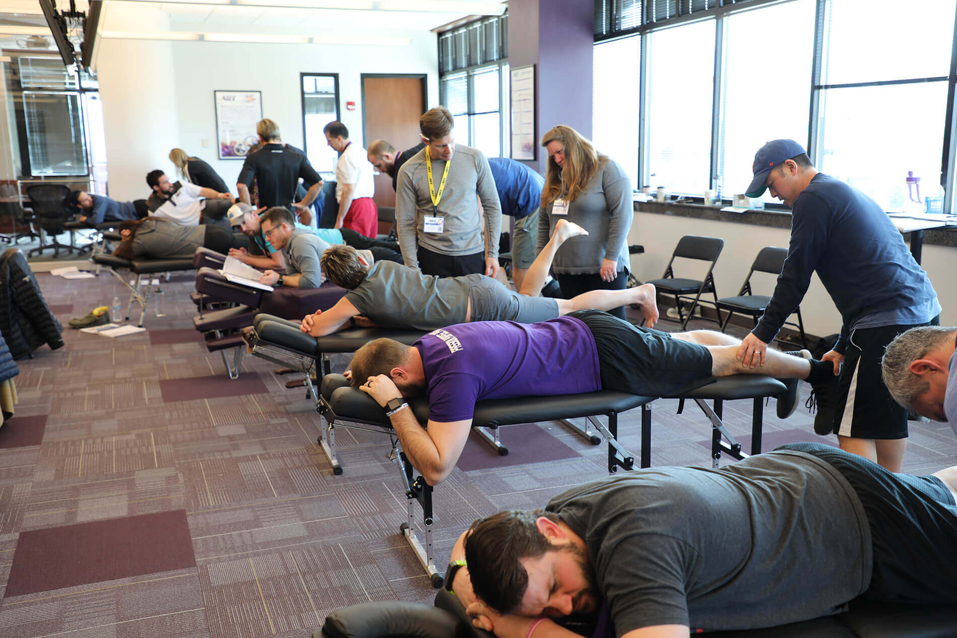
    Course Instruction & *Hands-on Practice
  • 10:00 – 10:15 am

    Break
  • 10:15 am – 12:00 pm

    Instruction Continues
  • 12:00 – 1:00 pm

    Lunch Break
  • 1:00 – 3:00 pm

    Instruction Continues
  • 3:00 – 3:15 pm

    Break
  • 3:15 – 5:00 pm

    Instruction Continues
Day 4

Sunday

Testing
  • 7:00 am – 12:00 pm

    Testing (20-minute increments)

Snacks will be provided during scheduled breaks. Providers should expect to receive test scores after the completion of the seminar, usually within 72 hours.

*Hands-on practice of treatment protocols: During the seminar, attendees practice protocols on one another and instructors to ensure they perform the appropriate movement, incorporating correct tension, touch, and palpation, for each protocol. Attendees practice asking open-ended questions, such as: nature of injury, duration of signs and symptoms; intensity of signs and symptoms; Initial onset of pain; aggravations to worsen pain; and pain classification (sharp, dull, achy, throbbing). Attendees are instructed to look for swelling, discoloration, visual patient discomfort, analyze gait/movement/posture as it relates to pain and the nervous system, all while actively palpating various structures.

Attendees learn to effectively palpate soft tissue structures, namely, nerves, muscles (including origin, insertion, muscle belly, and tissue abnormalities), ligaments, retinaculum, fascia, joints, and bony structures. While palpating, attendees are trained to ask the patient to perform active range of motion and describe symptoms, including burning, tingling, pain, and numbness, while the attendee actively feels for abnormalities of the structure. Attendees also learn to take the patient through passive range of motion while palpating structures and feeling for abnormalities in tissue structure, with a focus on nerves, while simultaneously soliciting patient feedback.

Protocols Covered
  • Digital Nerves at Palmar Fascia & Lumbricals
  • Median Nerve at Thenar Muscles
  • Median Nerve at Carpal Tunnel
  • Median Nerve at Pronator Teres / Flexor Digitorum Superficialis
  • Median Nerve at Ligament of Struthers
  • Median Nerve at Coracopectoral Tunnel
  • Median Nerve at Lower Scalenes
  • Superficial Branch Radial Nerve at Distal Brachioradialis
  • Superficial Branch Radial Nerve at Proximal Brachioradialis/Extensor Carpi Radialis Longus/Supinator
  • Radial Nerve (Deep Branch) at Proximal Wrist Extensors
  • Radial Nerve (Deep Branch) at Supinator
  • Radial Nerve at Brachioradialis/Brachiali
  • Radial Nerve at Lateral Intermuscular Septum
  • Radial Nerve at Triceps
  • Radial Nerve at Triangular Interval/Superior Radial Tunnel
  • Radial Nerve at the Coracopectoral Tunnel
  • Radial Nerve at Lower Scalenes
  • Ulnar Nerve at Hypothenar Muscles
  • Ulnar Nerve at Tunnel of Guyon
  • Ulnar Nerve at Pronator Quadratus
  • Ulnar Nerve at Flexor Carpi Ulnaris
  • Ulnar Nerve at Medial Intermuscular Septum
  • Ulnar Nerve/Medial Chord at Subscapularis
  • Ulnar Nerve at Lower Scalenes
  • Phrenic Nerve at Omohyoid
  • Brachial Plexus at Omohyoid
  • Nerve Roots at Upper Scalenes
  • Nerve Roots at Anterior Intertransversarii
  • Medial Antebrachial Cutaneous Nerve at Fascia
  • Lateral Antebrachial Cutaneous Nerve at Biceps/Brachialis
  • Inferior Lateral Brachial Cutaneous Nerve at Triceps
  • Musculocutaneous Nerve at Coracobrachialis
  • Axillary Nerve at Quadrangular Space
  • Medial Pectoral Nerve at Pectoralis Minor
  • Long Thoracic Nerve at Serratus Anterior
  • Greater Occipital Nerve at Nuchal Line/Upper Trapezius
  • Greater Occipital Nerve at Semispinalis Capitis
  • Third Occipital Nerve at Upper Trapezius
  • Suboccipital Nerve at Superior Oblique
  • Suboccipital Nerve at Rectus Capitis Major/Minor
  • Dural Motion
  • Isolation of Dural Entrapment
  • Dura at the Brainstem and Rectus Capitis Anterior
  • Dorsal Ramus of C2 at Inferior Oblique/Posterior Intertransverse Muscle
  • Dorsal Rami of C4-C8 at Trapezius/Splenius Capitis
  • Spinal Accessory, Vagus, and Facial Nerves at Digastric
  • Lesser Occipital Nerve at SCM/Splenius Capitis
  • Greater Auricular/Transverse Cervical/Spinal Accessory/Supraclavicular Nerve at Splenius Cervicis/Levator Scapulae/Posterior/Middle Scalene
  • Greater Auricular Nerve/Transverse Cervical Nerve at Sternocleidomastoid
  • Spinal Accessory Nerve at Sternocleidomastoid/Middle Scalene
  • Spinal Accessory Nerve at Trapezius/Rhomboids
  • Dorsal Scapular Nerve at Splenius Cervicis/Iliocostalis Cervicis
  • Dorsal Scapular Nerve at Rhomboids
  • Suprascapular Nerve at Suprascapular Foramen
  • Intercostal Nerves at Intercostals
  • Dorsal Rami T1-T6
  • Dorsal Rami T7-T12
  • Dorsal Rami L1-L3
  • Dorsal Rami S1-S3
  • Superior Gluteal Nerve at Gluteus Medius/Minimus
  • Posterior Cutaneous Nerve at Gluteus Maximus
  • Pudendal Nerve at Ischial Tuberosity
  • Pudendal Nerve at Pelvic Floor
  • Pudendal Nerve at Obturator Internus
  • Sciatic Nerve Roots at the Foramen
  • Sciatic Nerve at Hip Rotators
  • Sciatic Nerve at Long Head of Biceps Femoris
  • Tibial Nerve at Tendinous Arch of Soleus
  • Posterior Tibial Nerve in the Deep Compartment
  • Posterior Tibial Nerve at Tarsal Tunnel
  • Common Peroneal Nerve at Biceps Femoris
  • Common Peroneal Nerve at Peroneus Longus
  • Deep Peroneal Nerve at Tibialis Anterior
  • Deep Peroneal Nerve at Extensor Digitorum
  • Deep Peroneal Nerve at Extensor Hallucis Longus
  • Deep Peroneal Nerve at Extensor Retinaculum
  • Medial Calcaneal Nerve at the Flexor Retinaculum
  • Medial and Lateral Plantar Cutaneous Nerve at Plantar Fascia
  • Digital Nerves at Flexor Tendons/Lumbricals
  • Autonomic Chain at Psoas
  • Obturator Nerve at Adductor Brevis/Longus
  • Femoral Nerve Roots at the Foramen
  • Femoral Nerve at Psoas
  • Femoral Nerve at Psoas and Quadratus Lumborum
  • Lateral Femoral Cutaneous Nerve at Quadratus Lumborum/Psoas
  • Lateral Femoral Cutaneous Nerve at Inguinal Canal
  • Genitofemoral Nerve at Psoas
  • Femoral Nerve at Femoral Sheath
  • Saphenous Nerve at Adductor Canal
  • Saphenous Nerve at Sartorius / Gracilis
Nerve Certification Icon

The best and brightest in the field.

Image of 5 students/providers intently looking on during an ART seminar.

Still have questions?

Connect with our Seminars team.

  • This field is for validation purposes and should be left unchanged.